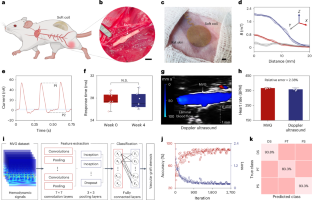
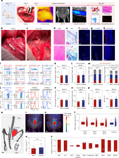
a, b, The rats with MVGs anastomosed to the femoral artery behaved and moved normally with no appreciable skin necrosis or swelling for up to 120 days post-implantation. Scale bars, 2 cm. c, The ultrasound probe is positioned above the femoral incision area to evaluate the patency of the anastomosed MVG. Partially created with BioRender. d, e, (d) The original Doppler ultrasound image alongside (e) the vascular color flow image captured the state of the anastomosed MVG two weeks post-surgery. f, g, (f) The original Doppler ultrasound image alongside (g) the vascular color flow image captured the state of the anastomosed MVG four weeks post-surgery. The use of color gradients in these images illustrates the blood flow, demonstrating the MVG’s patency during the early stages of recovery. These images provide critical insights into the graft’s immediate function and its integration within the host’s vascular system, showcasing the preservation of blood flow and the absence of apparent major obstructions. h, The photograph depicts the femoral incision area post-implantation, showcasing wound healing with no obvious signs of wound infection or inflammation. Scale bar, 1 cm. i, An infrared thermal image of the femoral incision area reveals an even temperature distribution. This uniform thermal profile is indicative of a healthy healing process, with no obvious signs of excessive heat that could denote infection or inflammation. Scale bar, 2 cm.
— Source: Nature Biotechnology (https://www.nature.com/articles/s41587-025-02619-7)